8/1/2019
The Lowdown on Leafy Gall
Melodie Putnam

Plant pathologists love pathogens the way growers love plants: the diversity of shapes and forms, the way the organisms can exploit certain environmental niches to grow and flourish, and the challenges of learning about unique types. The difference is that growers generally have a dim view of pathogens.
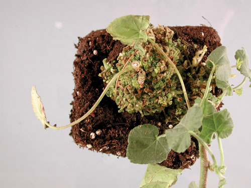
Pictured: A relatively large basal leafy gall of lavatera.
Bacterial diseases are difficult for plant health managers because the bacteria cannot be seen unless present in copious masses—and sometimes not even then. Leaf and stem spots, and vascular wilts are some of the more familiar manifestations of bacterial problems. But what about bacteria that are present on or in the plant for long periods before symptoms appear? Or when disease is completely overlooked when symptoms do show up because it isn’t recognized as anything significant until it’s too late? This is the situation presented by the bacteria known as Rhodococcus fascians.
These bacteria cause an unusual type of growth malformation, where an excess number of buds are produced. These buds don’t expand to their full potential, but remain in arrested development as either unexpanded buds or as buds that begin to grow, but are then stalled somewhere during the process. This aggregation of immature leaf or shoot buds is called a leafy gall.
In most plants, the leafy galls form at or near the soil surface, but they can appear anywhere on the plant, although I haven’t seen floral structures affected. There may be other symptoms as well—for example, lily bulbs may be elongated and twisted, and in some plants, fewer roots are produced—but generally the most frequent symptom noticed by most growers is leafy galls. Leafy galls can slide under your radar, as products used to promote branching may produce similar-looking structures. If in doubt, it would be prudent to have the tissue tested.
The bacteria appear to live on the plant surface for a time until some population size is reached or some other trigger occurs, after which the bacteria can penetrate deep into the interior of the plant. We were able to confirm this using electron microscopy. Rhodococcus doesn’t live in the vascular tissue, and hence, isn’t present throughout all parts of an infected plant. However, it can be present on or in plants without showing symptoms, which means taking cuttings from infected plants is a genuine risk: the cuttings may develop symptoms weeks—or even months in the case of woody plants—later. Even tissues up to five inches away from a basal leafy gall may carry the bacteria.
Leafy galls don’t appear to seriously disrupt the normal functioning of the affected plant and generally don’t lead to the plant’s death. I have kept several symptomatic campanula plants in a greenhouse for over 15 years and the plants are thriving in spite of the infection (we’ll get back to these plants later).
However, the dense structure of a cluster of half-expanded leaf buds can catch and retain water, leading to secondary decay by organisms like Botrytis. Leafy galls can get to be rather large (think melon sized) and are unattractive, and in essence, are a cosmetic problem. But in a business where beauty is the trade and conformation to type is the expectation, gross malformations lead to real and significant losses. Worse, diseased plants can act as a point source of infection, “sharing” their bacteria with nearby plants, leading to an epidemic of malformed plants that are unsellable. This occurs with particularly susceptible plants, such as Veronica spicata and Leucanthemum superbum cultivars.
Transmission
In addition to moving on infected propagation material, the bacteria are spread in water splash and can live for at least a few months in water. In the early 1970s researchers in California found indirect evidence of persistence of the bacteria in irrigation canals in which leucanthemum were growing, and in our own studies, we found the bacteria could be recovered from regular tap water, which we’d spiked with the bacteria, after three months. After that time, the bacterial populations declined to the point where they were undetectable.
Bacteria are resourceful little creatures, though, and we do know at least some strains of Rhodococcus can persist in a state of lowered metabolism under conditions of nutritional stress (e.g., starvation) and rebound when more favorable conditions are encountered. Since these bacteria don’t form any sort of spores or other structures that are resistant to environmental degradation, this suggests an adaptive mechanism for survival, allowing persistence under conditions that would kill other bacteria.
Rhodococcus isn’t known to be naturally vectored by insects, but transmission may occur on seeds of some plants, including petunia, and possibly nasturtium, argyranthemum, dianthus and pelargonium.
We investigated the possibility of transmission on cutting tools experimentally. We cut through a leafy gall on Oenothera speciosa and then sequentially cut through 10 healthy plants. We did this multiple times and didn’t see disease develop in any of the plants by the end of three months.
At the end of the experiment, we selected tissue from each plant, divided the tissue into two parts and used one part to culture from and the other to test for pathogenic Rhodococcus using a molecular test (PCR). Although the bacteria could be detected using PCR on the first plants cut, we didn’t recover any living bacteria. We obtained the same results when using petunia as the experimental plant. This suggests the bacteria aren’t readily transmitted on cutting tools.

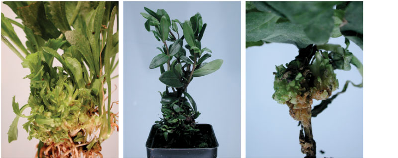
Pictured: • Clusters of shoots and leaves are typical of a leucanthemum leafy gall.
• Woody plants are also susceptible to pathogenic Rhodococcus, such as this hebe.
• An aerial leafy gall of petunia.
In nurseries with a Rhodococcus problem, where did it come from? There was some historical information that suggested the primary means the bacteria move within a nursery is by propagation from diseased plants, but what about the possibility of movement between nurseries? This was a question I was particularly interested in, and which I and my colleagues at Oregon State University took a look at.
I had a collection of R. fascians strains from multiple states and dozens of plant species obtained over the period of 15 years. I also had some from non-plant sources, such as from an ice core from Greenland. Through modern sequencing methods, all the genetic information contained within 60 different bacterial strains was determined.
We found that genetically similar bacteria (too closely related to be attributed to chance) were present at three or more nurseries, which suggests the bacteria were being moved between sites. We also found that I’d obtained genetically similar bacteria from one site in sampling periods that were four years apart, which indicates the bacteria were probably persisting at the site over those four years. And remember the campanula plant I mentioned earlier? I was able to recover pathogenic Rhodococcus 15 years after I first received it. Clearly, maintaining an individual stock plant for years increases the chances that it will eventually become diseased, and if infected by Rhodococcus, these plants can be a source of infection for multiple years.
Susceptible plants
Rhodococcus fascians has been found to affect a large number of plant species. There are an even greater number of plants that are susceptible when intentionally treated with the bacteria, but these plants don’t become infected without being inoculated. For example, digitalis will become diseased if treated with the bacteria, but in over 30 years of diagnosing diseases, I’ve never seen a Rhodococcus-infected digitalis plant, nor have I found any scientific publications that mention natural infection of this host.
A study we undertook showed there were genetic differences between strains of the bacteria, but it appears to be the species of plant that has more influence on how severe the disease will be. We found no correlation between strains of the bacteria and the plant species from which the bacteria were recovered.
Up until 2014, Rhodococcus fascians was known as the only species of the genus capable of causing plant disease, although it was also found in some pretty interesting places, such oceans, flies that frequent sores on sheep and in caves. One result of our genetic analysis of the isolates we examined was that those that cause disease in plants consisted of 13 genetically distinct groups of bacterial strains. That is, the strains that are capable of causing the growth malformations that are typical of a “Rhodococcus fascians” infection are not just one species, but could be differentiated into at least 13 groups different enough to be separate species. To simplify things, we decided to refer to strains that cause disease in plants as pathogenic Rhodococcus.
Disease management
We’ve looked at various control compounds that are registered for use against bacteria to see if any were effective in controlling the bacteria. In multiple assays using bacteria in culture, ZeroTol 2.0 and Oxiphos were superior to the 10 other products we tested. However, neither ZeroTol 2.0 nor Oxiphos were able to prevent disease on inoculated plants. The concentrations of bacteria we used in these experiments were high in order to get sufficient infection to see a difference between treatments, and it may be that the products would be effective at lower bacterial concentrations. However, there’s no cure once a plant is infected. And since the bacteria may be inside the plant, topical applications may not be effective regardless of the concentration of bacteria on the surface.
Once diseased plants are observed, get laboratory confirmation, since not all things that produce Rhodococcus-like malformations are due to Rhodococcus.
As of this writing, there are no quick assays that growers can perform themselves, although two companies—Loewe (www.loewe-info.com) and Lifeasible (www.lifeasible.com)—sell materials to perform enzyme-linked immunosorbent assays (ELISA), which are more suitable to laboratory testing than field testing. These assays will detect any Rhodococcus “fascians” and will not discriminate between those that are pathogenic and those that are not, which is why I do not recommend their use. A detection of Rhodococcus that isn’t pathogenic could result in needless destruction of crops.
If a pathogenic Rhodococcus infection has been confirmed, strict sanitation is the primary means of disease management. I recommend destroying all affected plants so the bacteria cannot spread within your facility. Whether or not neighboring plants become diseased depends on the inherent susceptibility of the species. Oenothera, petunia, veronica and leucanthemum are among the most likely to pick up an infection when close to diseased plants of another species. Pots, flats and surfaces on which diseased plants have been in contact should be sanitized with bleach or a quaternary ammonium product. I’ve sampled and haven’t been able to recover live bacteria from a nursery’s landscape fabric that was bleached after an infection had been found.
To prevent new introductions of disease, segregate newly obtained susceptible cuttings or plants, especially those four mentioned above. Try to source a supplier of clean plants. Unfortunately, I’ve diagnosed tissue culture plants with pathogenic Rhodococcus, so obtaining plants grown from tissue culture doesn’t ensure freedom from this pathogen. GT
Melodie Putnam is the Director of the Oregon State University Plant Clinic and has been working with Rhodococcus “fascians” for nearly 20 years. She may be reached at putnamm@Oregonstate.edu or at her Twitter account: @OR_PlantClinic. The Plant Clinic web site is: bpp.oregonstate.edu/plant-clinic.